This page covers inspection of the eye and fundoscopy (ophthalmoscopy). A complete examination of the eye should include assessment of the optic nerve (vision), eye movement and reflexes. These are covered in the cranial nerve examination page (cranial nerves II – VI).
Introduction
Introduce yourself and confirm the patient's name and date of birth. Explain the procedure and obtain consent. Position the patient on a chair and ask them to remove their glasses or contact lenses before proceeding to wash your hands.
There is a lot to explain in a fundoscopy examination. Keep your language simple and concise. For example:
"I will be using this instrument which has a magnifying glass to try to see the back of your eye. I will need to come close to your eye and shine a light. This can be a little uncomfortable but not painful. If at any point it gets too uncomfortable and you want to pause or stop, please just let me know."
If a patient has poor eyesight, ask them what their corrections are, as you will need to adjust the numbered wheel on the fundoscope to correct for refractive errors. The negative numbers (red) correct myopia (short-sightedness). The positive numbers (green) correct hypermetropia (long-sightedness).
General inspection
Ask the patient if they are in any pain and are comfortable. Look briefly around the bed and observe the face and eyes for any surgical scars, obvious discharge, erythema (suggesting inflammation) or swelling. Causes of such swelling include, but are not limited to infection, allergy and trauma.
Closer inspection
Ask the patient to look at a fixed point in the distance. Use the light of the fundoscopy to closely inspect the external eye, looking at the sclera, conjunctiva, cornea, pupil and iris.
Observe the sclera for a red appearance, which signifies dilation of ocular vessels. This is commmonly caused by conjunctivitis, but, with the persistent presence of pain, can signpost more serious pathologies including anterior uveitis, keratitis and acute closed-angle glaucoma. If a red sclera is accompanied by significant discomfort and irritation, suspect a corneal abrasion.
Inspect the eyeball for any foreign bodies such as a contact lens fragment. Closely look for any scarring which may indicate to a corneal abrasion. Check for any deposits or ulcers which may be present in patients with anterior uveitis.
Finally note any discharge. Clear and wet discharge is usually physiological or due to allergy, whereas sticky discharge is an indicator of infection and is common in infectious conjunctivitis, orbital cellulitis and blepharitis.
Conjunctivitis is inflammation of the conjunctiva, a heavily vascularised epithelial layer covering the sclera. There are many types of conjunctivitis, and the presentation varies with each. Eye redness, excessive tears and “puffy eyes” are three features common to all.
Allergic conjunctivitis: Allergens such as pollen may cause irritation to the conjunctiva. The patient will have itchy eyes, a clear, watery discharge and other symptoms of allergy such a nasal congestion.
Viral conjunctivitis: Caused by viruses such as adenovirus and herpes simplex virus. The patient will have flu-like symptoms and there may be a sticky clear discharge from the eye.
Bacterial conjunctivitis: Caused by bacteria such as Staphylococcus and Streptococcus. The patient will typically have a rapid onset of symptoms a sticky, swollen eyelid often with a “gritty” sensation, and a sticky discharge, usually yellow in colour.
Irritant conjunctivitis: Various acidic and alkaline chemicals in day-to-day products such as shampoo can cause temporary inflammation of the conjunctiva, resulting in itchy eyes and crusting of the eyelashes.
Other causes of conjunctivitis involved autoimmune phenomena such as in reactive arthritis.
Uveitis is inflammation of the uvea, an eye structure composed of the iris, ciliary body and choroid. Depending on the structures affected, this can be further subdivided into posterior and anterior uveitis.
The patient with present with blurred vision, eye redness, pain and photophobia. If the posterior uvea is affected there may also be floaters or peripheral visual field loss. In some cases of longstanding anterior uveitis, the patient has a small, irregular pupil due to adhesions of the iris to the lens, and with severe inflammation there may be a hypopyon (a yellow proteinous exudate in the anterior chamber of the eye) or keratic precipitates, the latter of which can be viewed with a slit-lamp.
Causes of uveitis are plentiful and include infectious causes such as herpes simplex virus and syphilis, and non-infectious causes such as Behçet's disease, sarcoidosis and systemic lupus erythematosus.
Keratitis is inflammation of the cornea. The patient will present with eye redness, sudden eye pain, intense photophobia and impaired eyesight. It may be caused by viruses such as herpes simplex virus, which often leaves a pathopneumonic dendritic corneal ulcer, bacteria such as Staphylococcus, fungi, amoebae and parasites.
Glaucoma is an optic neuropathy in which there is visual field loss. Two main types include closed-angle and open-angle glaucoma. These are defined by the presence of ocular hypertension (increased pressure in the eye) which compresses on the optic nerve. Whilst causes are usually primary in origin, these glaucoma types may also be due to trauma, uveitis or steroids.
Closed-angle glaucoma: The angle between the iris and cornea narrows, abruptly blocking the trabecular meshwork which drains aqueous humour from the anterior chamber of the eyeball. This results in sudden ocular hypertension, and as such there is a rapid onset of symptoms including a painful red eye, impaired vision and halos around lights. Often the pressure will result in a fixed, dilated pupil that is minimally responsive to light and cupping of the optic disc.
Open-angle glaucoma: This is the most common cause of glaucoma. The trabecular meshwork becomes blocked over time due to risk factors such as age, ethnicity and myopia. This results in a progressive ocular hypertension and is mostly asymptomatic. Over time there may be a peripheral visual field loss and on examination you may see cupping of the optic disc.
There is also a third type of glaucoma called normal- or low-tension glaucoma. Low vascular perfusion of the optic nerve makes it more susceptible to the normal pressures of the eye leading to damage of the nerve. The presentation is similar to that of traditional open-angle glaucoma.
Trauma to the cornea frequently results in abrasions. Regular causes of trauma are contact lens insertion/removal, scratching of the eye and dirt in the eye.
Orbital cellulitis describes inflammation of the tissues posterior to the orbital septum, a membranous fascia which forms the anterior boundary of the orbit. This is a medical emergency. Inflammation of tissues anterior to this boundary is known as periorbital cellulitis. Whilst not an emergency, it must still be managed in hospital as it may progress to orbital cellulitis.
Patients will present with swelling of the eye and surrounding areas, erythema around the eye and discharge. Orbital cellulitis will additionally present with a number of red flag signs: proptosis, opthalmoplegia, pain on eye movement and loss of vision.
The causes of each are bacterial in nature and are usually Staphylococci or Streptococci.
Blepharitis is inflammation of the eyelids. This may be infective or allergic in nature. The patient will have burning, itchy eyes and a visible swollen and erythematous eyelid. Crusting may be visible on the lid margins.
Before you commence examination with the fundoscope you must prepare your patient and the instrument. Darken the room.
Pupil dilation
Ask the patient if they have received any eye drops. Ideally, the patient should have received mydriatic medication (e.g. tropicamide) prior to fundoscopy in order to maintain pupil dilation.
Tropicamide (1%), an anti-muscarinic drug used to dilate the pupil, generates short acting mydriasis (pupillary dilation) and cycloplegia (paralysis of the ciliary muscles, consequently causing a loss of the accommodation reflex). The process starts to occur after 5 minutes, takes up to 30 minutes to reach full effect and lasts for about 2 – 3 hours. It is contraindicated in patients with glaucoma, as the drug may increase intraocular pressure. Side effects of the drug include blurriness and double vision.
Choice of beam
Check the fundoscope is functioning suitably and choose the white beam of an appropriate size using the aperture dial. Most ophthalmoscopes have various sizes of beams – small, medium and large:
The small beam is used when a pupil is constricted, e.g. in a bright room.
The large beam can be utilised if a dilating agent (e.g. tropicamide) has been used.
The medium beam is appropriate in a dark room when a dilating agent hasn’t been used.
Next, use the rheostat the adjust the brightness such that the pupil is minimally constricted.
Pre-adjustment for refractive error
Adjust the dioptre wheel of the fundoscope to correct for your refractive error. For example, if you are short sighted with an error of -2, then adjust the wheel of the fundoscope to -2, which is normally coloured red.
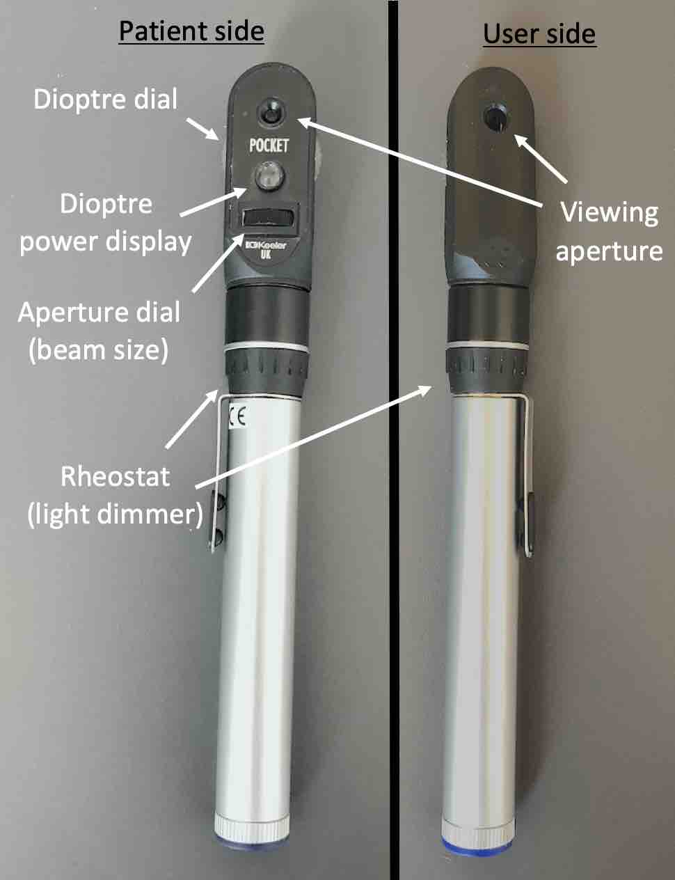
Parts of the fundoscope.
The red reflex
Begin the fundoscopy examination in the patient’s “good” eye. Request the patient to focus on a point in the distance.
Now depending on which eye you are examining first, hold the fundoscope with your right hand (to examine the patient’s right eye) or your left hand (to examine the left eye). Look through the scope approximately 50cm away from the patient’s eye and shine the light on their pupil. Inspect for a red reflection; this is a red/orange colour seen through the scope which occurs due to reflection of light from the patient’s retina. Continue to look for a red reflection as you bring the fundoscope in medially towards the nose while still keeping the light on the pupil a distance of 50cm; moving nasally ensures checking of the optic disc.
If there is an abnormal, white reflection (leukocoria) at any point, this is suggestive of pathology. In adults it is usually due to cataracts. In children it may indicate the presence of congenital cataracts, a retinoblastoma or retinopathy of prematurity.
An absence of a red reflex or darkness suggests a retinal detachment or glass eye.
Currently we are only assessing one eye. Once you have finished examining the fundus however, we will need to repeat the entire process in the other eye also.
A cataract describes a progressive clouding of the lens in the eye, ultimately leading to blurriness of vision. The most common risk factor is age, but its onset can be accelerated by both diabetes mellitus and hypertension. Some chromosomal diseases and infections in-utero can also lead to congenital cataracts which are present at birth.
This is a malignant cancer of the retina frequently caused by mutation to the tumour suppressor gene RB1. It will be present at birth. The patient will be blind in that eye and will have no red reflex.
Also known as retrolental fibroplasia, retinopathy of prematurity is a disease which occurs as a result of oxygen therapy use in premature babies. This can affect development of the retina resulting in scarring, and in worst cases, retinal detachment. As such, babies will not present with a red reflex.
A retinal detachment occurs when the outer pigment epithelium detaches from the inner retina which contains the neurons. Patients will present with photopsia (flashes of light) and a dramatic increase in visual floaters. There will be visual field loss, starting peripherally and then eventually moving centrally. On fundoscopy, you will see a grey, lifeless retina which bulges forward.
Take your free hand, that is, the hand that is not currently holding the fundoscope, and place your index finger on the patient’s brow. Starting from 50cm away, look through the fundoscope and move it in towards the pupil until you can visualise the back of the eye.
If you are examining the patient’s right eye, then you will be using your right hand to hold the fundoscope. Therefore, you will rest your left index finger on the patient’s right brow. The reason for doing this is to ensure that you do not move too close to the patient when examining the fundus.
Adjustment for refractive error
At this point, you may need to correct the fundoscope for refractive error once again, this time to adjust to the patient’s error, until you have a clear image of the back of the eye. If the patient is short-sighted, turn the wheel towards the negative, and if long-sighted, then turn it towards the positive.
Some individuals choose not to pre-adjust the fundoscope and instead simply start with a setting of +8 and continue turn the wheel down until they get a clear image.
The optic disc, macula and retinal vessels on the fundus.
Optic disc
Find a blood vessel and track its progress in until you find the optic disc. To do this, follow it in the direction in which the vessel amalgamates with others, forming fewer and fewer vessels. Once at the disc, identify its characteristics, specifically the margin, colour, contour and cup-to-disc ratio. A normal disc will be pink with a well-defined margin, a pale central physiological cup and a cup-to-disc ratio between 0.3 and 0.5:
Margin: An indistinct margin can indicate optic disc swelling. This may be due to numerous causes including as glaucoma, optic neuropathy, retinal vessel occlusion and raised intracranial pressure. When it is caused by the latter, it is known as papilloedema.
Colour: A pale optic disc signifies that there has been atrophy due to the death retinal ganglion cell axons. Risk factors include glaucoma, ischaemic optic neuropathy and optic neuritis.
Contour: The border of the disc is normally well demarcated and clear. If the disc is swollen, then a raised contour will be visible.
Cup-to-disc ratio: This is the diameter of the cup in relation to the optic disc. For example, if the cup takes up 3/10 of the disc the cup-to-disc ratio is 0.3. A ratio above 0.5 alludes to glaucoma, while an absence of a cup implies papilloedema.
An optic neuropathy describes damage to the optic nerve. Glaucoma, a major cause, is detailed in a complex box earlier in the page.
Optic neuropathy can also occur when there is ischaemia (ischaemic optic neuropathy) and inflammation (optic neuritis). The causes and presentations of each of these vary as follows:
Ischaemic optic neuropathy: Common causes include diabetes mellitus, hypertension and temporal arteritis. The patient will often have sudden loss of vision, and will present with a pale, and oedematous optic disc with poor margins on examination.
Optic neuritis: Causes include multiple sclerosis, diabetes mellitus, syphilis and ethambutol. The patient will present with pain, blurred vision and a deterioration in visual acuity and colour vision.
Retinal artery occlusion is an emergency condition in which there is occlusion of a central of branch retinal artery. It may be caused by thrombus formation, arteritis or emboli.
Patients will present with a sudden loss of vision, which may be temporary (amaurosis fugax) if caused by an embolus, or long-standing if caused by thrombus formation or arteritis. On fundoscopy, the arteries will be narrow and truncated. Fluid leakage will lead to a swollen retina which is pale and oedematous with a cherry red spot on the macula. .
Retinal vein occlusion involves occlusion of a central or branch retinal vein. Risk factors for its development include diabetes mellitus, hypertension, glaucoma and hyperviscosity syndromes.
Patients will present with reduced visual acuity. On fundoscopy there will be numerous flame haemorrhages in the distribution of the vein and cotton wool spots at areas of retinal ischaemia. The optic disc will be swollen with poor margins and a raised contour due to fluid leakage.
After assessing the disc, move on the assessing the retina and its vessels. Examine the four quadrants of the retina methodically in a clockwise or anticlockwise method. The retinal vessels will radiate out from the centre of the optic disc, the veins appearing thicker than the arteries. If there is an abnormally large number of vessels then this suggests that there has been neovascularisation, which occurs in diabetes mellitus, retinopathy of prematurity and retinal vein occlusion.
As you examine the quadrants, look closely for the following signs:
Arteriolar narrowing: Some arteries may have a narrowed, silver or copper wire appearance. These phenomena occur in grade 1 hypertensive retinopathy.
Arteriovenous (AV) nicking: When an artery crosses a vein, it may “nick” the vein resulting in compression. This is visible as bulge on either side of the crossing. AV nicking may be seen in grade 2 hypertensive retinopathy.
“Cotton-wool” spots: These fluffy-white spots on the retina occur as a result of ischaemia. They are commonly present in grade 3 hypertensive retinopathy and diabetic retinopathy.
Exudates: Lipid residues from vessels that have leaked or haemorrhaged will form yellow deposits known as hard exudates. Typical causes include grade 3 hypertensive retinopathy, diabetic retinopathy and retinal vein occlusion.
Next, observe for the presence of any haemorrhages. Bleeding in the retina can be due to a multitude of causes each resulting in its own haemorrhagic pattern. Haemorrhages can be subdivided by their appearance:
Dot-and-blot haemorrhages: These will appear as small red dots or blots anywhere on the retina. They are a result of microaneurysms which have haemorrhaged. They are most commonly associated with diabetic retinopathy.
Flame haemorrhages: These are larger, red marks with a “feathered-” or “flame-like” appearance. They suggest a larger bleed, and may be present in grade 3 hypertensive retinopathy, normal-tension glaucoma and retinal vein occlusion. The latter in particular will result in multiple flame haemorrhages appearing.
Large haemorrhages: Very large bleeds, for example due to retinal trauma or vessel tearing in retinal detachment will result in visibly large red or pigmented marks which cause visual field loss.
Finally, observe for any uncommon abnormalities such as white, round photocoagulation spots which occur following laser treatment for diabetic retinopathy, or rarer black, pigmented spicules (a minute sharp-pointed object) in the periphery, which are present in retinitis pigmentosa.
Hypertension can damage the retinal arteries leading to hypertensive retinopathy. This will present with blurred vision and headaches as the disease progresses. There will also be a number of changes which can be seen on fundoscopy. These ocular manifestations can be classified by Keith-Wagener-Barker grades.
Grade 1: Identified by the presence of arteriolar narrowing only. This may be present as copper or silver wiring.
Grade 2: Identified by the presence of arteriolar narrowing in addition to arteriovenous nicking.
Grade 3: Identified by the features of grade 2 with any of these additional features: retinal oedema, cotton wool spots, hard exudates and flame haemorrhages.
Grade 4: Identified by the features of grade 3 plus papilloedema.
Hyperglycaemia in diabetes mellitus can cause damage to the retinal arteries causes diabetic retinopathy. This is one of the world’s leading causes of blindness. Often there are no symptoms early on, but as the disease develops the patient will eventually present with blurred vision. Fundoscopy can be used to identify the disease in its early stages, and the ocular manifestations vary with the stage of the disease:
Non-proliferative diabetic retinopathy (NPDR): In the mild stage, NPDR will present with dot haemorrhages which develop due to the emergence of microaneurysms. In moderate NPDR you may see the emergence of blot haemorrhages, venous engorgement and hard exudates. Over time, ischaemia will result in the development of cotton wool spots. Once these features affect all four quadrants the retinopathy is considered severe.
Proliferative diabetic retinopathy (PDR): This is a progression of NPDR and can have any of its features. Continued ischaemia will eventually result in neovascularisation; new vessels grow out of the retina and into the vitreous humour.
Diabetic maculopathy: If there is lipid deposition in the macula (hard exudates) or there is macular oedema due to leaky blood vessels, this is described as diabetic maculopathy. This is independent of the non-proliferative/proliferative nature of the retinopathy.
Retinitis pigmentosa is an autosomal inherited disease in which black, pigmented spicules develop in the periphery of the retina leading to progressive blindness. This starts peripherally, progresses to “tunnel-vision” and then eventually complete blindness.
Request the patient to look directly at the light of your fundoscope very briefly to examine the macula.
Identify any exudates; diabetes retinopathy can sometime affect the macula only. Look for the normal pink colour macula. A pigmented macula may indicate age related macular degeneration.
Age-related macular degeneration (ARMD) is the most common cause of blindness in the United Kingdom. As the macula is the centre of the retina, the loss of vision is also central. Risk factors include age, being female, smoking and a family history of the condition.
There are two major types. In nonexudative (dry) and exudative (wet) ARMD:
Nonexudative ARMD: This develops gradually and without pain. On fundoscopy you may see hard exudates in the macula and surrounding areas. There may also be pigment released from dying cells.
Exudative ARMD: This develops suddenly and has rapid progression. On fundoscopy there will be neovasculariation and exudative oedema. This needs to be treated urgently.
Remember to repeat all the steps from examining the red reflex to the fundus in the patient’s other eye using your other hand to hold the ophthalmoscope.
Completion
Thank the patient and wash your hands. Summarise your findings and present to the examiner. Offering appropriate differential diagnoses, relevant further examinations/tests, such as a full cranial nerve assessment, and if appropriate, capillary glucose, a urine dipstick, blood pressure and retinal photography.
Interactive Markscheme
When assessing each other, click on each list item as you go along. Doing so will turn the list item green. Make careful note of any steps missed at the end.
We recommend completing any examination or procedure in under 10 minutes, but you can adjust the timer to suit your needs.
:
This Interactive Markscheme is for the fundoscopy (ophthalmoscopy) examination only. A complete examination of the eye would involve assessment of vision, eye movement and reflexes, all of which can be found in steps 7-18 of the cranial nerve examination list.
Introduction: “Hello, I’m SimpleOSCE and I am a medical student. I’ve been asked to examine the back of your eyes today; would that be okay? Can I confirm your name and DOB? Thank you.”
Explain what you are going to do: ““I will be using this instrument which has a magnifying glass to try to see the back of your eye. I will need to come close to your eye and shine a light. This can be a little uncomfortable but not painful. If at any point it gets too uncomfortable and you want to pause or stop, please just let me know.”
Wash your hands.
Ask about pain and discomfort.
Inspect the face and eyes for any scars, discharge, erythema or swelling.
Closely inspect the external eye for redness, foreign bodies, scars, deposits, ulcers and discharge.
Darken the room.
Ask the patient if they have had their pupils dilated.
Check the fundoscope is functioning suitably and choose the appropriate beam.
Optional: Adjust the wheel correcting for refractive error.
Request the patient concentrate on a point in the distance.
Optional: Start the fundoscopy with the “good” eye.
Holds fundoscope correctly, examining the right eye with right hand and left eye with left hand.
Inspect for a red reflex in that eye.
Bring the light in nasally to check the optic disc.
Optional: Adjust the wheel of the fundoscope again to adjust for the patient’s refractive error.
Examine the optic disc (margin, colour, contour and cup-to-disc ratio).
Examines four quadrants of the retina systematically.
Request the patient to look into light and examine the macula.
Repeat steps 13-19 in the other eye.
Wash your hands and thank the patient.
"To conclude, I would like to take a history a perform a full cranial nerve examination.”